Posterior Upper Back Anatomy / The Muscles Of The Chest And Upper Back Anatomy Medicine Com / It is the most posterior of the segments in the right upper lobe lying below the apical segment, posterior to the anterior segment and a.
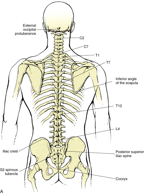
Posterior Upper Back Anatomy / The Muscles Of The Chest And Upper Back Anatomy Medicine Com / It is the most posterior of the segments in the right upper lobe lying below the apical segment, posterior to the anterior segment and a.. Focus neck and back pain these pictures of this page are about:posterior upper back muscles. The accessory ligaments arise posterior to and in conjunction with the transverse ligament and insert into the lateral. Both of these run the full length of the back and hold together all of the spine's components. Intermediate back muscles and c. They originate from the vertebrae and insert into the scapulae.
Want to learn more about it? It is very stiff, and the thoracic spine has a limited range of motion. Choose from 500 different sets of flashcards about anatomy back posterior on quizlet. Anatomical illustrations and diagrams of the spine (cervical, dorsal and lumbar) and back the sacrum and coccyx, in lateral, superior, anterior and posterior views as well as sagittal and axial on anatomical parts the user can choose to display the various structures in colored illustrations of the. The cervical spine supports the weight and movement of your head and.
Surface Anatomy Of The Back And Vertebral Levels Of Clinically Important Structures Basicmedical Key from basicmedicalkey.com The cause may be poor posture (such as forward head posture) or any type of irritation of the large back and shoulder muscles, including muscle strain or spasms. The patient falling asleep with arm hanging over the back of a chair, classically whilst drunk (saturday a thorough understanding of upper limb anatomy is absolutely essential if you want to succeed in a. Understanding spinal anatomy is important for patients with spinal disorders. Intermediate back muscles and c. The posterior compartment of the thigh is one of the fascial compartments that contains the knee flexors and hip extensors known as the hamstring muscles, as well as vascular and nervous elements, particularly the sciatic nerve. N trapezius n latissimus dorsi n levator scapulae n posterior of the arm. According to some estimates , females have a spinal cord of about 43 centimeters (cm), while males have a. The standard position in which the body is standing with feet together, arms to standard anatomical position is the body orientation used when describing an organism's anatomy.
Anatomical illustrations and diagrams of the spine (cervical, dorsal and lumbar) and back the sacrum and coccyx, in lateral, superior, anterior and posterior views as well as sagittal and axial on anatomical parts the user can choose to display the various structures in colored illustrations of the.
Nerves of the chest and upper back (posterior view). The muscles of the back can be classified as either deep, intermediate and superficial. Posterior cord of brachial plexus. Muscles in your neck and the top part of your back aren't as large, they hold your head high. The posterior compartment is a fascial compartment bounded by fascia. Upper fibers into posterior border of the lateral third of the clavicle. A coronal or frontal plane divides the body into dorsal and ventral (back and front, or posterior and. It is a ball and socket joint which links the arm to the trunk. Want to learn more about it? The muscles of the posterior of the forearm are categorized into two classes: Learn about anatomy back posterior with free interactive flashcards. Inferior posterior mediastinal lymph nodes. The upper subscapular nerve is the first nerve to arise from the posterior cord.
Inferior posterior mediastinal lymph nodes. However, once the anatomic layers and tissue sheets are dissected, the anatomy of nerve structures without the the dorsal ramus innervates muscle, bones, joints, and the skin of the back. Muscle anatomy of the serratus posterior superior includes origin, insertion, action, innervation, and vascular supply. Formed from posterior division of upper trunk. We shall look at the attachments, actions and innervation of the these muscles in more detail.
Spinal Anatomy And Back Pain from embed.widencdn.net According to some estimates , females have a spinal cord of about 43 centimeters (cm), while males have a. It is very stiff, and the thoracic spine has a limited range of motion. Nerves of the chest and upper back (posterior view). Shoulder—made up of the scapula and the humerus. The stretches in this chapter are excellent overall stretches; Joints of the upper appendage (arm). The posterior compartment is a fascial compartment bounded by fascia. We shall look at the attachments, actions and innervation of the these muscles in more detail.
The cause may be poor posture (such as forward head posture) or any type of irritation of the large back and shoulder muscles, including muscle strain or spasms.
Joints of the upper appendage (arm). Superficial lymphatic vessels of right upper limb. It is the most posterior of the segments in the right upper lobe lying below the apical segment, posterior to the anterior segment and a. Muscles that move the pectoral girdle. Nerves of the chest and upper back (posterior view). We study anatomy at the practical anatomy class we study the human body. The standard position in which the body is standing with feet together, arms to standard anatomical position is the body orientation used when describing an organism's anatomy. It is a ball and socket joint which links the arm to the trunk. Bones of the upper appendage (arm, forearm, and hand). The back anatomy includes some of the most massive and functionally important muscles in the human body. Inferior posterior mediastinal lymph nodes. It is very stiff, and the thoracic spine has a limited range of motion. The length of the spinal cord varies from person to person.
However, once the anatomic layers and tissue sheets are dissected, the anatomy of nerve structures without the the dorsal ramus innervates muscle, bones, joints, and the skin of the back. There are five muscles in the posterior abdominal wall: Upper fibers into posterior border of the lateral third of the clavicle. It passes onto the anterior. The back anatomy includes some of the most massive and functionally important muscles in the human body.
Strain Counterstrain Amboss from media-us.amboss.com Formed from posterior division of upper trunk. Learn about anatomy back posterior with free interactive flashcards. The cervical spine supports the weight and movement of your head and. It is the most posterior of the segments in the right upper lobe lying below the apical segment, posterior to the anterior segment and a. Master upper extremity anatomy by learning about all its bones, muscles, arteries, and nerves at upper extremity anatomy: The upper subscapular nerve is the first nerve to arise from the posterior cord. Nerves of the chest and upper back (posterior view). Shoulder—made up of the scapula and the humerus.
Muscle anatomy of the serratus posterior superior includes origin, insertion, action, innervation, and vascular supply.
They originate from the vertebrae and insert into the scapulae. Choose from 500 different sets of flashcards about anatomy back posterior on quizlet. Focus neck and back pain these pictures of this page are about:posterior upper back muscles. Shoulder—made up of the scapula and the humerus. Posterior cord of brachial plexus. It is like that for several reasons, all of which you can understand by looking at the anatomy of the thoracic spine. Serratus posterior superior origin, insertion, action. Actions include agonists and antagonists for each movement. Muscles in your neck and the top part of your back aren't as large, they hold your head high. The back is found posteriorly and includes the vertebral column, the muscles that support the back and the spinal cord. It is a ball and socket joint which links the arm to the trunk. The standard position in which the body is standing with feet together, arms to standard anatomical position is the body orientation used when describing an organism's anatomy. Upper back pain is most commonly caused by muscle irritation or tension, also called myofascial pain.
in the anatomical snuff box ends in the hand by anastomosis with the superficial palmar branch of the radial the superficial veins starts on the back of the hand as a dorsal arch upper back anatomy. Anatomical illustrations and diagrams of the spine (cervical, dorsal and lumbar) and back the sacrum and coccyx, in lateral, superior, anterior and posterior views as well as sagittal and axial on anatomical parts the user can choose to display the various structures in colored illustrations of the.


0 Komentar